Sanionia nivalis
Ann. Bot. Fenn. 26: 411, f igs. 10 - –12. 1989.
Plants medium-sized to large. Stems unbranched, sometimes sparsely and irregularly branched, or rarely ± pinnate. Stem leaves falcate or strongly falcate, rarely straight or almost so, not plicate, plicate, or rarely strongly plicate, 0.5–1.7 mm wide; base ovate-triangular to broadly ovate; margins plane or sometimes partly recurved distally, strongly denticulate distally; apex acuminate to long-acuminate; costa in bottom of shallow, wide-angled fold (or not in fold); alar region ± isodiametric, transition to supra-alar cells gradual, alar and supra-alar cells together often in ± homogeneous ovate basal marginal group, supra-alar cells rectangular or long-rectangular, ± echlorophyllose, walls thin, eporose, region equal in size to or much larger than alar region; apical laminal cells with distal ends occasionally prorate abaxially. Perichaetia with inner leaves ± suddenly narrowed to apex, margins strongly denticulate to dentate distally, apex acute or short-acuminate. Capsule ± erect; exothecial cells ± isodiametric, in (1–)2–3 rows; exostome specialized, teeth long, or apical end of distal tooth portion somewhat reduced, narrow distally, border not widened at transitional zone in pattern of external tooth; endostome specialized, in recently dehisced capsules hyaline or slightly yellowish, basal membrane constituting 17–33% endostome height, processes and basal membrane with one or both cell wall layers strongly perforated all over, cilia rudimentary or absent.
Habitat: Large late snow beds, shores of glacier-fed brooks
Elevation: low to high elevations
Distribution

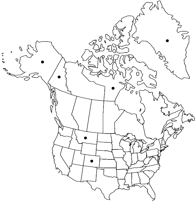
Greenland, Nunavut, Yukon, Alaska, Colo., Mont., n Europe.
Discussion
Sanionia nivalis is easily separated from the other three species of Sanionia by its reduced and irregularly perforate endostome, the structure of the alar and supra-alar cells, and the acute to short-acuminate inner perichaetial leaves with strongly denticulate or dentate distal margins; the other three species have long-acuminate inner perichaetial leaves with finely denticulate or denticulate distal margins. In addition, the stem leaves of S. nivalis are more short-acuminate and have more strongly denticulate margins than in the three other species.
Selected References
None.